
Organ-on-a-chip technology to reduce side effects of radiotherapy
By Candy Gibson
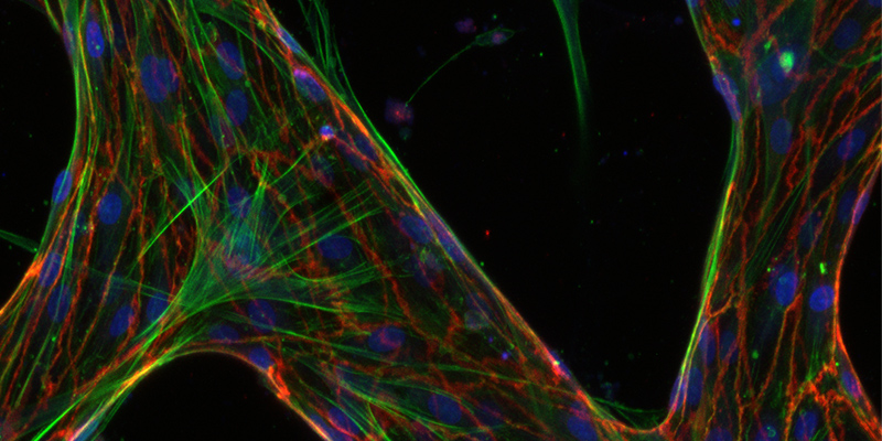 HEALTH The microvasculature formed inside the chip. Photo by Joe Vittorio.
The debilitating side effects of radiotherapy could soon be a thing of the past thanks to a breakthrough by UniSA and Harvard University researchers.
UniSA biomedical engineer Professor Benjamin Thierry is leading an international study using organ-on-a-chip technology to develop 3D models to test the effects of different levels and types of radiation.
A microfluidic cell culture chip closely mimics the structure and function of small blood vessels within a disposable device the size of a glass slide, allowing researchers and clinicians to investigate the impact of radiotherapy on the body’s tissues.
To date, scientists have relied on testing radiotherapy on cells in a two-dimensional environment on a slide.
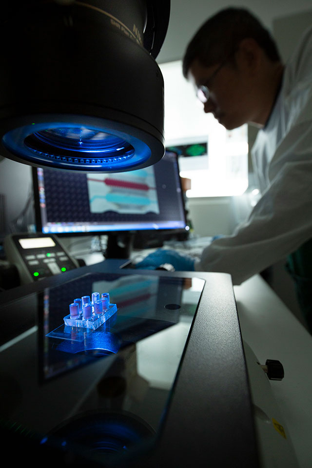 Microfluidic cell culture chip: UniSA bioengineer Dr Chih-Tsung Yang, the co-first author of the study, pictured with the microfluidic cell culture chip in the foreground. Photo by Joe Vittorio.
Professor Thierry, from UniSA’s Future Industries Institute (FII) and the ARC Centre of Excellence in Convergent Bio-Nano Science and Technology (CBNS), says the organ-on-a-chip technology could reduce the need for animal studies and irrelevant invitro work, both of which have major limitations.
“An important finding of the study is that endothelial cells grown in the standard 2D culture are significantly more radiosensitive than cells in the 3D vascular network. This is significant because we need to balance the effect of radiation on tumour tissues while preserving healthy ones,” Prof Thierry says.
The findings, published in Advanced Materials Technologies, will allow researchers to fully investigate how radiation impacts on blood vessels and – soon – all other sensitive organs.
“The human microvasculature (blood vessel systems within organs) is particularly sensitive to radiotherapy and the model used in this study could potentially lead to more effective therapies with fewer side effects for cancer patients,” Prof Thierry says.
More than half of all cancer patients receive radiotherapy at least once in the course of their treatment. While it cures many cancers, the side effects can be brutal and sometimes lead to acute organ failure and long-term cardiovascular disease.
Prof Thierry’s team, including UniSA FII colleague Dr Chih-Tsung Yang and PhD student Zhaobin Guo, are working in close collaboration with the Royal Adelaide Hospital and Harvard University’s Dana-Farber Cancer Institute with the support of the Australian National Fabrication Facility.
“Better understanding the effect of radiotherapy on blood vessels within organs – and more generally on healthy tissues – is important, especially where extremely high doses and types of radiation are used,” Dr Yang says.
The researchers’ next step is to develop body-on-chip models that mimic the key organs relevant to a specific cancer type.
Other Stories
- Painless needles on the way for vaccinations
- Hi. How can I help you? Health clinic embraces the robot age
- Virtual health coach ‘Paola’ will help you get fit and eat well
- Carbon-free renewable energy solution to ‘heat up’ industry
- From the Vice Chancellor
- Achievements and Announcements
- The technology to take a jury into a 3D crime scene
- Oscars 2019 plays it safe with Green Book, lacks enlightened thinking
- Organ-on-a-chip technology to reduce side effects of radiotherapy
- Find out about UniSA’s dedicated student support services (Video)
- International defence research lab with France planned for Adelaide
- New innovation hub in Whyalla to nurture regional startups
- The latest books from UniSA researchers
- In Pictures