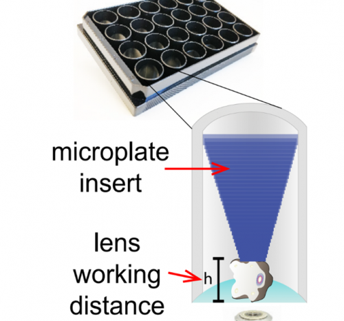
Microplate inserts for growing organoids in a fixed location.

Benefits
- Easy integration into existing cell culture platforms and automation systems.
- Ideally suited for advanced microscopy and imaging experiments.
- Cost-effective approach allowing scalability.
Background
Organoids are “mini-organs” that are grown within a lab using cell culture techniques and have emerged as a useful tool to study the development of human tissues, organs and disease.
Organoids have been shown to more accturately reproduce the human genetics and tissue organ organisation of the organ/disease they model, and can be used as a preclinical platform to analyse patient response to treatment.
Organoids have the potential to reduce the reliance on in vivo testing (animal models), which are expensive and limited in representing human tissue and disease development.
The global 3D cell culture market is expected to reach ~$4b annually, growing at nearly 30% annually (Allied Market Research).
Adoption of organoid cell cultures for clinical and pathological testing remains nascent. This is due to limitations in the capacity to image them at the whole organoid level and at high-resolution.
Current methods for organoid culture use constant agitation and generate tissues that range from 0.5 to 2 mm of size.
These culture conditions (agitation) and size (>1 mm for mature organoids) are not compatible with most inverted microscopes already available in most of research laboratories. Therefore, there is an urgent need for methods that allow the culture and growth of organoids that permit whole organoid live cell imaging and analysis over different times scales (minutes to weeks). They also need to be compatible with time scales for organoid development and permit the monitoring of organoid response to drug treatment.
Technology
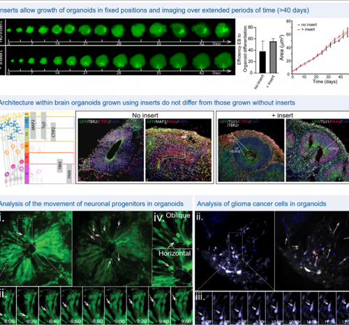
The microplate inserts have been designed to be adaptable to standard multi-well plates that can be used to grow multiple organoids in pre-defined and fixed XYZ coordinates. This technology facilitates high-resolution imaging of whole organoids, allowing precise assessment of organoid growth and morphology, as well as cell tracking within the organoids, over long periods of time (minutes to weeks).
The technology has been demonstrated by tracking neocortex development through neuronal progenitors in brain organoids, as well as the movement of patient-derived glioblastoma stem cells within healthy brain organoids.
This new bioengineering platform constitutes a significant advance that permits long term detailed analysis of whole organoids using multimodal inverted fluorescence microscopy.
Potential applications
Laboratory consumables for R&D.
Biotechnology and Pharmaceutical companies.
Cell culture companies.
IP Status
The technology is protected by a provisional patent.
Opportunities
Licensing and/or co-development.